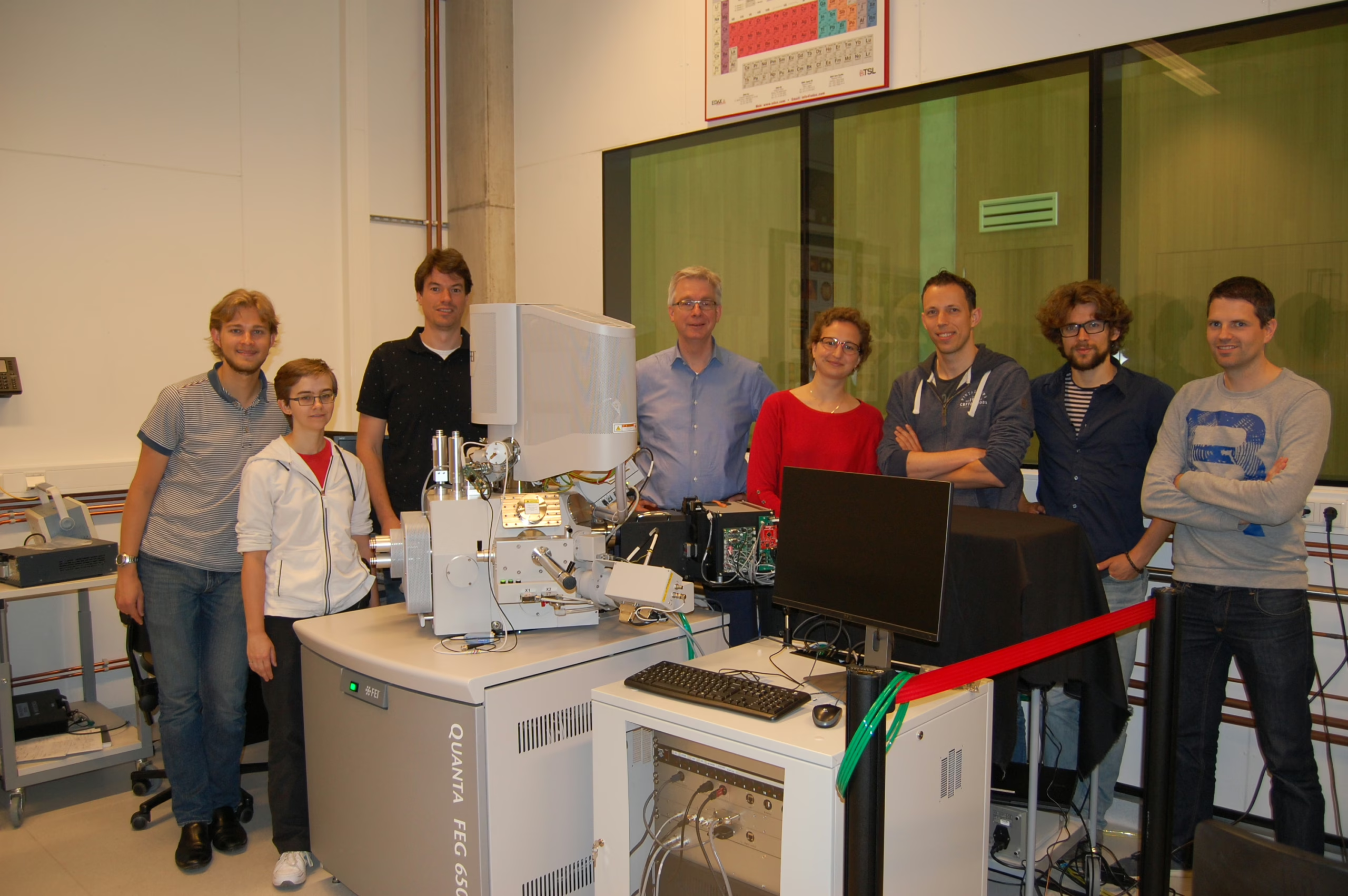
During a festive celebration on June 22, AMOLF has officially opened a new cathodoluminescence microscope in Albert Polman’s group that enables ultrafast optical measurements with a spatial resolution of 10 nanometer.
The new microscope has been built as part of a collaboration between AMOLF, FEI and Delmic. Together, these partners will investigate the potential of time-resolved cathodoluminescence microscopy as a new characterization tool in nanophotonics, quantum optics, semiconductor opto-electronics, biology, geology, photovoltaics, photocatalysis, and other areas.
The new instrument integrates (1) a FEI Quanta 650 scanning electron microscope (SEM) equipped with an ultrafast beam blanker, (2) electronics hardware developed at AMOLF that drives the beam blanker, (3) a Delmic SPARC cathodoluminescence imaging system that collects and analyzes the optical emission induced by the electron beam, and (4) single-photon counting/correlation and time-resolved spectroscopy assembled at AMOLF. The new instrument delivers electron pulses with a duration down to 1-5 ns enabling the combination of ultra-fast fluorescence lifetime imaging with nanoscale spatial resolution, and quantum optical studies in a broad range of systems.
In a further advanced design (to be realized later this year), the SEM electron cathode will be driven by a 250-fs pulsed UV laser beam, creating (single) electron pulses with a duration of 1 picosecond. This will enable ultrafast pump-probe imaging spectroscopy with a spatial resolution of 10 nm. The research program on the new microscope is funded by an ERC Advanced Investigator Grant that was awarded to Polman in 2016.
Cathodoluminescence imaging spectroscopy has been developed over the years at AMOLF as a nanophotonics measurement technique by three subsequent PhD students: Ernst Jan Vesseur, Toon Coenen and Benjamin Brenny, in addition to several postdocs and master students and many technical specialists at AMOLF. Over 40 scientific papers have been published so far, and are listed on http://www.erbium.nl/arcis.htm.